Researchers make waves in the ultrasound world
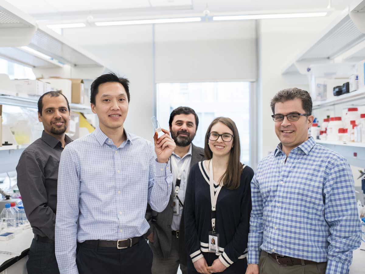
Photo: Scott Tsai demonstrating the microfluidic device developed by research team. From left: Vaskar Gnyawali, Professors Scott Tsai and Raffi Karshafian, Jennifer Kieda and Professor Michael Kolios (not pictured: Byeong-Ui Moon). Photo by Carrie Duncan.
A team of Ryerson researchers, led by Scott Tsai, have developed a new method to create the uniformly minuscule microbubbles most desirable for use in ultrasound imaging.
The cross-disciplinary team, based out of the iBEST laboratories at St. Michael’s Hospital, includes Tsai from Ryerson’s Mechanical and Industrial Engineering department and Raffi Karshafian and Michael Kolios from the Physics department. Co-supervised graduate student Vaskar Gnyawali and postdoctoral fellow Byeong-Ui Moon created the models and conducted the experiments. The project is being lauded as a simple and elegant solution to shrinking microbubbles for imaging and therapeutic uses. Their paper Honey, I shrunk the bubbles: microfluidic vacuum shrinkage of lipid-stabilized microbubbles was published in the Soft Matter journal of the Royal Society of Chemistry (external link) . The project was supported by funding from the Natural Sciences and Engineering Council of Canada (NSERC) Discovery and Engage grants.
Current techniques for producing the microbubbles, involving shaking liquids containing the surfactants (substances designed to reduce the surface tension of liquids), for bubbles, don’t provide the desired uniformity of size required by researchers specializing in ultrasound.
“We had tried before to produce microbubbles using microfluidics, but we couldn’t get them small enough,” said Tsai. “We just needed this simple breakthrough to get the size down.”
The team’s research addresses this problem by using a microfluidic chamber that employs vacuum microchannels to pull air from the bubbles as they pass through a serpentine channel, shrinking them down by pulling air through the channel wall between the vacuum and the bubble suspension. The channel wall material is non-permeable to water but allows the passage of gas (air) removed by the vacuum. The results are stable, uniform bubbles of ideal size for ultrasound use. Previous attempts at making bubbles through microfluidics depended on being able to manufacture small enough channels to shrink the bubbles, and these were unsuccessful at producing the tiny bubbles necessary for ultrasound imaging or therapy.
According to Kolios, this use of microbubbles can be fine-tuned by employing mono-disperse (or same-sized) bubbles. “At two microns,” said Kolios, “they resonate. They produce a lot of ultrasound signal.”
Raffi Karshafian noted the potential for loading the bubbles with drugs to target delivery to specific areas in the body. “By exerting stress on the bubbles using ultrasound, we can cause inertial cavitation (popping) of the bubbles,” he said. “With bubbles that are the same size, we can predict that they will behave the same way and we can enhance accumulation and transportation of drug molecules such as chemotherapy drugs.”
The next challenge for the team will be to understand the physics behind the pressure needed for the shrinkage, to better control the size of the bubbles and to scale up production of the bubbles, as the current system would require up to a year to produce enough bubbles for one clinical procedure. Tsai’s goal is to reduce that time to about one hour. He is currently putting together a proposal for the next phase of development, which would allow him to scale up the project to produce the bubbles at a faster rate.