Research
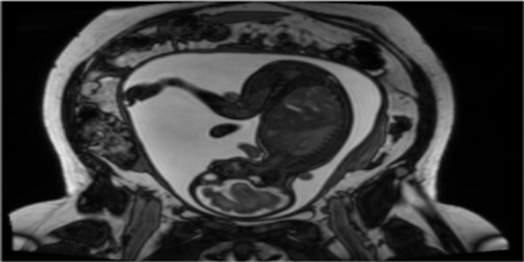
At the MFI Laboratory, we develop and use a variety of magnetic resonance (MR) sequences that facilitate non-invasive acquisition of physiological data of the mother and her fetus. We then apply these techniques to understand how maternal lifestyle choices and gestational disorders alter maternal and fetal physiology, fetal development and consequently, postnatal health.
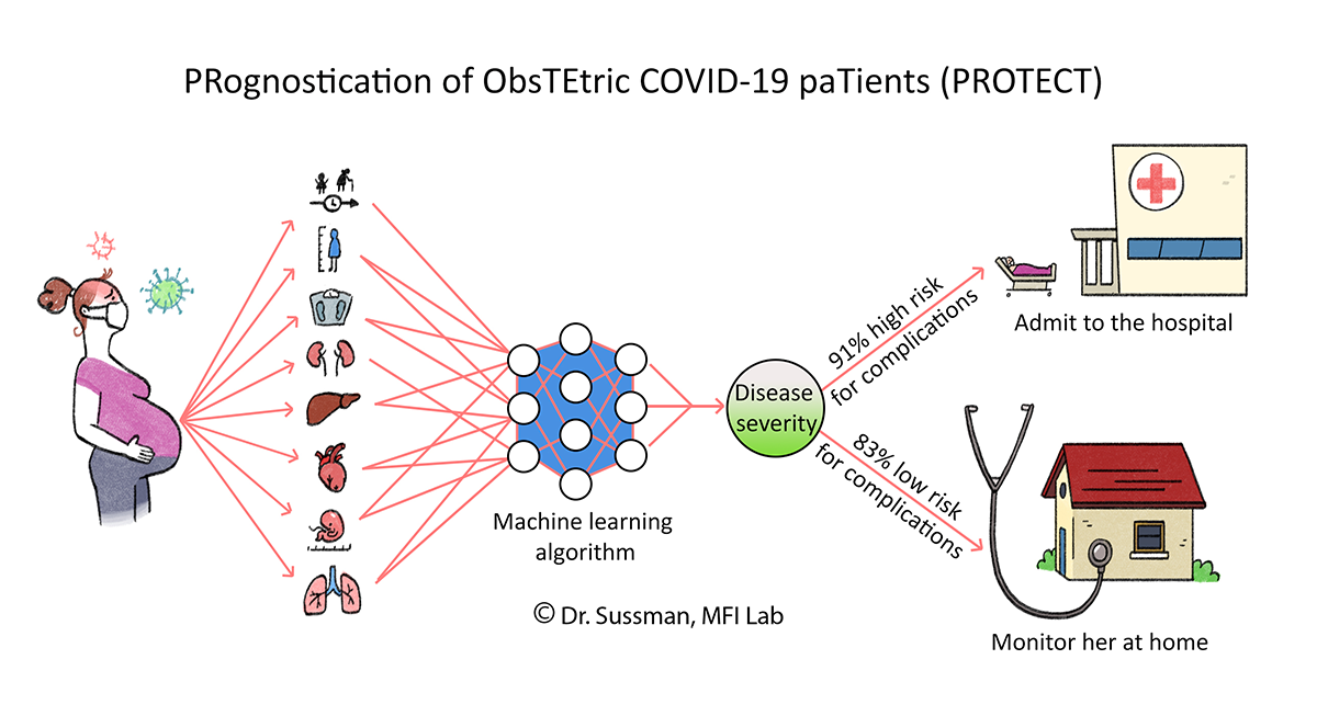
Prognostication of Pregnant COVID-19 Patients
Through an international collaboration, we are creating an artificial intelligence algorithm that identifies patterns and predicts disease severity and progression in pregnant women with COVID-19.
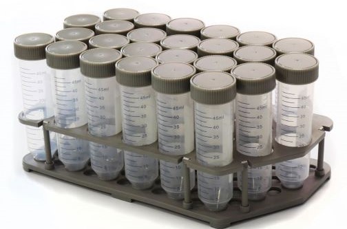
Tissue Mimicking Materials
We are synthesizing novel materials that mimic the MRI properties of different human tissues.
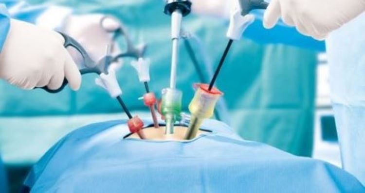
Surgical Skills
We are developing new techniques for medical learners to develop and practice their surgical skills and receive constructive feedback from their educators.
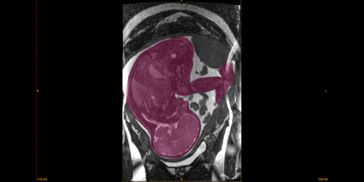
Automatic Fetal Image Segmentation and Classification
We are using machine learning approaches along with image processing techniques to develop novel algorithms for segmentation and disease classification of the placenta, fetus, and fetal organs from MR images. These will permit earlier and more accurate disease detection and facilitate interventions.
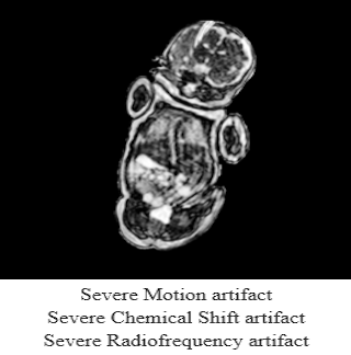
Quality Assurance and Artifact Correction
We are employing machine learning techniques to carry out automatic quality assurance of medical images. This includes assessment of artifact severity and removal of artifacts from images.
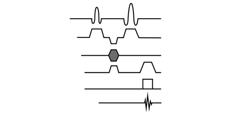
Imaging Fetal Glucose Metabolism
We are developing a novel MRI technique that will non-invasively assess glucose metabolism in fetal organs. This technique, called Chemical Exchange Saturation Transfer (glucoCEST), creates image contrast by exploiting magnetization transfer between glucose and surrounding water. This method will facilitate early diagnosis and treatment of fetal metabolic disorders and intra-uterine growth restriction.
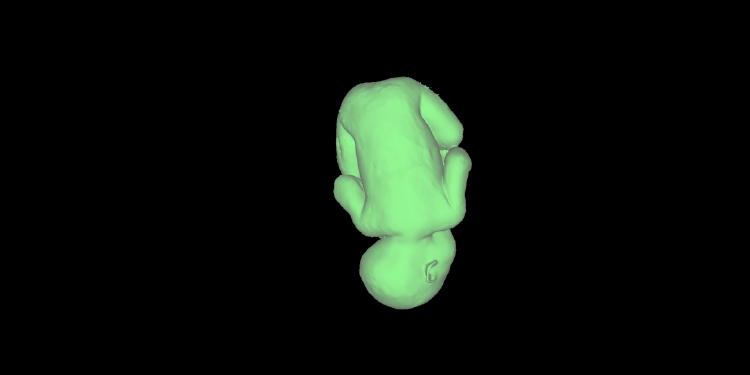
Fetal Phantom Synthesis
We are synthesizing 3D phantoms of the human fetus for magnetic resonance testing. This phantom will enable us to optimize various MR sequences as well as motion correction algorithms.
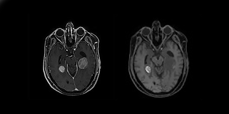
Smart Surveillance of Brain Tumours
Clinical surveillance of brain tumours is typically a manual task aimed at assessing tumour growth and transformation. We are working closely with neurosurgeons and radiologists to develop an algorithm that automatically carries out tumour grading from non-contrast enhanced 3D MRIs.
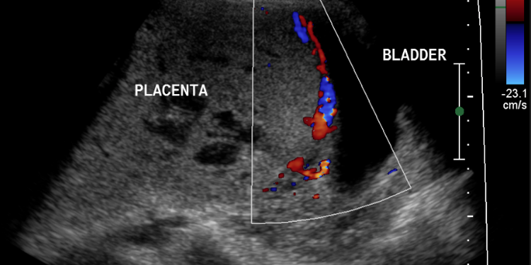
Invasive Placenta Identification and Classification
We are developing machine learning algorithms for detecting and classifying placenta accreta spectrum (PAS) disorder, where the placenta invades the myometrium. This will allow for earlier and more accurate diagnosis, facilitating clinical management and decreasing mortality.
Fetal Presentation Classification
We are utilizing machine learning to identify and classify fetal presentation and placental position from 3D MRIs in order to facilitate clinical management during delivery.