How to program a better tomorrow: Harnessing disruptive technologies
Innovation Issue 38: Summer 2023
Brain health: Using AI in medical imaging to improve patient outcomes
Idea to Innovation
Brain health: Using AI in medical imaging to improve patient outcomes
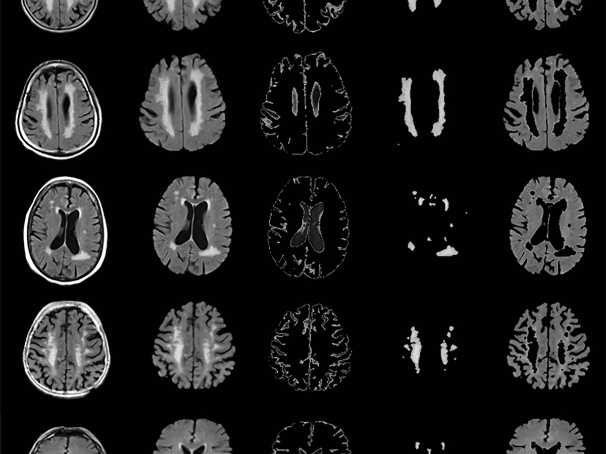
Professor April Khademi developed machine learning tools to automatically analyze MRI images and detect structures in the brain. Photo courtesy of April Khademi.
Research that began during Toronto Metropolitan University (TMU) professor April Khademi’s PhD has resulted in a patented tool that can help transform how clinicians diagnose dementia and vascular disease, leading to earlier detection and improved patient outcomes.
“Dementia and cerebrovascular disease place huge burdens on patients, their families and the healthcare system,” said the electrical, computer and biomedical engineering professor. “New technologies and tools that can diagnose patients earlier and more quickly can stratify patients into homogeneous subgroups for more personalized therapy.”
To diagnose and better understand these diseases, clinicians currently analyze MRI images of the brain and provide a qualitative description of what they see. Professor Khademi has developed machine learning tools that bring more automation and accuracy to this process. She also recently patented a normalization scheme used during the images’ pre-processing stage that allows for more consistency in their analysis and removes variability across different MRI scanners.
The team pre-processed over 400,000 MRI images from more than 100 health centres to prepare them for biomarker extraction. They also designed several types of segmentation tools that can automatically detect various structures in the brain, such as white matter, lesions, ventricles and the different hemispheres, in MRI images. These tools can quickly identify and extract biomarkers such as volume, size and shape that indicate specific biological processes, both normal and abnormal. The biomarkers are correlated with clinically relevant metrics to show if, for example, a patient has cognitive impairment or not.
Professor Khademi’s team successfully correlated blood biomarkers from patients who live with confirmed dementia. They are also beginning to gain unique insights that show the difference in pathology between Alzheimer's and cerebrovascular disease, which can help improve patient stratification and therapy selection.
“It also helps us understand a little bit about the etiology of disease, or the cause of disease,” said professor Khademi. “This is important because there are not a lot of therapeutic interventions for dementia and Alzheimer's disease. If we understand how the disease develops, maybe we can stand a chance to apply treatment earlier.”
Professor Khademi’s research has been developed in consultation with radiologists and neuroradiologists as well as a neurologist and a neuropsychiatrist. The next step in the research is to begin clinical translation, where radiologists will test the tools in practice.
Aside from detecting and diagnosing brain disease, these tools can help clinicians with robust, non-invasive disease monitoring for their patients. The technology can also be applied to other diseases in the future.
“I really believe that AI will change the way that medicine is practiced,” said professor Khademi. “Imaging is one of the most important tools used to manage patients. Imagine having a software-based toolbox that allows clinicians to get more objective measures of disease more easily.”
There are not a lot of therapeutic interventions for dementia and Alzheimer's disease. If we understand how the disease develops, maybe we can stand a chance to apply treatment earlier.

Read “Alzheimer's and Vascular Disease Classification using Regional Texture Biomarkers in FLAIR MRI (external link, opens in new window) ” in Neuroimage: Clinical.
Read “FLAIR MRI Biomarkers of the Normal Appearing Brain Matter are Related to Cognition (external link, opens in new window) ” in Neuroimage: Clinical.
Read “Segmentation of White Matter Lesions in Multicentre FLAIR MRI (external link, opens in new window) ” in Neuroimage: Reports.
Professor Khademi’s research is supported by the Natural Sciences and Engineering Research Council of Canada, the Alzheimer's Society of Canada, the government of Ontario, the Canadian Cancer Society and MITACs.