TMU researchers make breakthrough on diagnosing breast cancer with AI
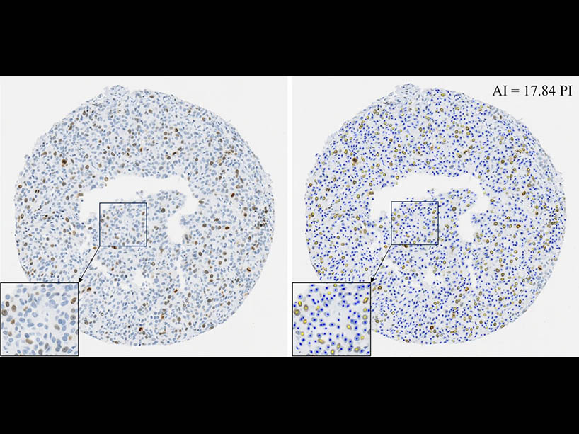
This figure from professor April Khademi’s published paper, AI improves accuracy, agreement and efficiency of pathologists for Ki67 assessments in breast cancer (external link) , shows tissue microarrays without the aid of AI (left) and with the aid of AI (right) showing the clarity the tool provides
Groundbreaking new research from TMU is showing remarkable success in using AI tools to assess and interpret breast cancer tissue, particularly focusing on the Ki-67 biomarker.
The TMU team developed a custom AI tool that helps pathologists make more consistent and reliable treatment decisions which can revolutionize cancer care. The research is led by professor April Khademi, Canada Research Chair and medical image analysis expert with support from PhD student Amanda Dy.
"This research represents a significant milestone in the integration of AI technologies into pathology workflows,” said Khademi, a biomedical engineer who was named Canada Research Chair in AI for Medical Imaging this year. “Findings suggest that AI tools not only improve assessment accuracy which results in more reliable treatment options, but also offer a substantial return on investment by streamlining workflow and enhancing patient care."
The Ki-67 biomarker, identified as a crucial indicator for deciding treatment, (external link) has traditionally posed challenges for pathologists - in that there is a high level of disagreement on how to score the tissue. This impedes the pathologists’ ability to deliver accurate and efficient assessments (external link) . Khademi’s pioneering research has been supported by a consortium of leading organizations including the Canadian Cancer Society (CCS) and the Vector Institute has propelled the field forward.
The AI-powered tool was rigorously tested and out-performed commercial solutions (external link) . This tool was tested live in an innovative study with 90 international pathologists and showed AI assistance significantly enhanced pathologists' assessments, leading to more accurate and efficient scoring of the Ki67 biomarker. Most notably, the AI tool improved pathologist scoring accuracy, agreement and turn-around-time.
“It's fundamental for engineers to collaborate and seek feedback from clinicians when designing and evaluating AI tools for clinical decision support,” said Dy. “This allows us to refine our work based on expert feedback while clinicians can grow more confident in the AI outputs,” adding that the key to growing confidence and trust in AI within the medical community lies in transparency and interdisciplinary collaboration.
Pictured here, PhD student Amanda Dy.
This study demonstrates how using AI can result in more consistent and reliable treatment decisions which has the potential to dramatically improve quality of care. The study, which is one of the largest of its kind, highlights the transformative potential of AI in pathology.
Khademi and Dy aim to leverage these findings for further advancements in cancer research and diagnostics.
"Our current study primarily focuses on the assessment stage, and our ultimate goal is to enhance treatment planning and patient outcomes," said Khademi. "By harnessing the power of AI, we aim to empower pathologists with additional insights and solutions that may have previously gone unnoticed, ultimately prioritizing the well-being of patients."
This groundbreaking research solidifies TMU’s position as a leader in health-care education and underscores Canada's prominence in the global scientific community. With continued collaboration and innovation, Khademi and her team are poised to revolutionize cancer diagnostics and improve patient care worldwide.
Related stories: